Deigram Of Outside Leg Muscles / Muscles Of The Leg And Foot Classic Human Anatomy In Motion The Artist S Guide To The Dynamics Of Figure Drawing - Diagramme schnell und einfach erstellen.
Deigram Of Outside Leg Muscles / Muscles Of The Leg And Foot Classic Human Anatomy In Motion The Artist S Guide To The Dynamics Of Figure Drawing - Diagramme schnell und einfach erstellen.. Your leg muscles are some of the hardest working muscles in your body. Leg pain can also be caused by blood clots, varicose veins or poor circulation. It contains the peroneus longus and peroneus brevis muscles. They receive their innervation via the deep. Rotating your upper leg and pelvis to the inside or outside of your body's center line.
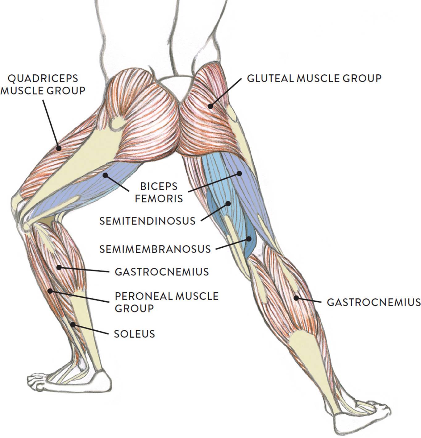
The muscles in this area move your leg out to the side. The muscles that make up the quadriceps are the strongest and leanest of all muscles in the body. The gastrocnemius is the larger calf muscle, forming the bulge visible beneath the skin. It is also visible on the medial edge of the thigh from the anterior. This muscular system chart shows in detail the deep layers of muscle on the back side of your body.
Muscular System Anatomy Lateral Leg Region Muscles Model Description Somso Youtube from i.ytimg.com On the outside of the thigh, this is the largest of. There are three parts to the trapezius. The hip muscles work together to carry out 4 different types of movement: Muscles and bones act together to form levers. They receive their innervation via the deep. The lower part of the trapezius ascends and depresses the scapula, while the transverse or middle region of the trapezius is what retracts the. This muscle runs along the outside of the back of your thigh and attaches to the top of the fibula (the smaller of the two bones of your lower leg). However, many reflex pathways are also active in the legs and foot.
The nerve signals in these reflexes come from stretch receptors located in the joints, ligaments, tendons, and even the muscles themselves.
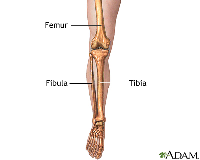
This muscle runs along the outside of the back of your thigh and attaches to the top of the fibula (the smaller of the two bones of your lower leg). The compartments are divided by septa formed from the fascia. Your leg muscles are some of the hardest working muscles in your body. Notice the upper leg has a biceps muscle just like the upper arm does. Muscles and bones act together to form levers. The anterior is located in the front portion of the leg. They also help with pointing the foot, or plantarflexion. This muscular system chart shows in detail the deep layers of muscle on the back side of your body. Keep in mind, while muscle cramps in the lower leg can last anywhere from a few seconds to several minutes, muscle soreness may continue for. This is the biggest muscle that is in the tibialis anterior. More specifically, this beautifully illustrated anatomy chart includes neck and shoulders, multiple views of the back and spine, and frontal views of each muscular extremity of the human body. Extension, flexion, adduction, and abduction. On the outside of the thigh, this is the largest of the four quadriceps muscles.
The four muscles that make up the quadriceps are the strongest and leanest of all muscles in the body.these muscles at the front of the thigh are the major extensors (help to extend the leg. We'll break down the anatomy and function of the upper leg, knee, lower leg. Most leg pain results from wear and tear, overuse, or injuries in joints or bones or in muscles, ligaments, tendons or other soft tissues. Muscles and bones act together to form levers. Anterior muscles of the lower leg and their functions.
Muscles Of The Leg And Foot Classic Human Anatomy In Motion The Artist S Guide To The Dynamics Of Figure Drawing from doctorlib.info Pain in your calf or thigh can be caused by muscle cramps, a pulled or strained muscle, or issues related to your nerves. The leg muscles diagram, will point out if the issue is with any tissue or with the bone. Biceps femoris (long head) biceps femoris (short head) semitendinosus. The compartments are divided by septa formed from the fascia. A muscle strain is a stretch or tear of muscle fibers. Reflexes help to maintain proper muscle tone, balance, and responsiveness of the legs and feet to stimuli such as stepping on a sharp object. However, many reflex pathways are also active in the legs and foot. Leg pain can also be caused by blood clots, varicose veins or poor circulation.
Notice the upper leg has a biceps muscle just like the upper arm does.
Extension, flexion, adduction, and abduction. It extends from the top of the femur at the hip and to the kneecap. The muscles that make up the quadriceps are the strongest and leanest of all muscles in the body. This muscular system chart shows in detail the deep layers of muscle on the back side of your body. This is why you have to indicate which biceps you are taking about when discussing one or other of these muscles. The fascial compartments of the leg are the four fascial compartments that separate and contain the muscles of the lower leg (from the knee to the ankle). On the outside of the thigh, this is the largest of. Observe the leg muscle diagram posted above and notice that there are many parts in the muscles.the largest muscle masses in the leg are present in the thigh and the calf. It gets its blood flow from the arteries in the tiberial artery. Add a frame to any. The muscles of the lower leg, called simply the leg by anatomists, largely move the foot and toes. Some common causes of leg pain include: The lower part of the trapezius ascends and depresses the scapula, while the transverse or middle region of the trapezius is what retracts the.
The anterior is located in the front portion of the leg. The lateral compartment is along the outside of the lower leg. A muscle strain is a stretch or tear of muscle fibers. It is also visible on the medial edge of the thigh from the anterior. Diagramme schnell und einfach erstellen.
Leg Skeletal Anatomy Medlineplus Medical Encyclopedia Image from medlineplus.gov Biceps femoris (long head) biceps femoris (short head) semitendinosus. The fascial compartments of the leg are the four fascial compartments that separate and contain the muscles of the lower leg (from the knee to the ankle). Other muscles run from your pelvis to your upper thigh bone. The lower part of the trapezius ascends and depresses the scapula, while the transverse or middle region of the trapezius is what retracts the. However, many reflex pathways are also active in the legs and foot. It contains the peroneus longus and peroneus brevis muscles. Tibialis anterior, extensor digitorum longus, extensor hallicus longus, fibularis (peroneus) longus, fibu. They allow you to move and provide support for your upper body.
In the leg, muscle strains happen when a muscle is either stretched beyond its limits or forced into extreme contraction.
On the outside of the thigh, this is the largest of the four quadriceps muscles. The muscles of the hip can be divided into three different. The muscles in the hip are responsible for the movement of the hip and, by proxy, the leg. Tibialis anterior, extensor hallucis longus, extensor digitorum longus, and fibularis tertius. The leg muscles diagram, will point out if the issue is with any tissue or with the bone. Add a frame to any. In the leg, muscle strains happen when a muscle is either stretched beyond its limits or forced into extreme contraction. More specifically, this beautifully illustrated anatomy chart includes neck and shoulders, multiple views of the back and spine, and frontal views of each muscular extremity of the human body. Some common causes of leg pain include: The hamstring muscle attachment points. Oleh riosandi444 agustus 05, 2021 posting komentar Anterior muscles of the lower leg and their functions. The muscles work together to enable movement and keep the hip in alignment.


Posting Komentar
0 Komentar